Pulse Ultrasound.
Specialist Vascular Ultrasound
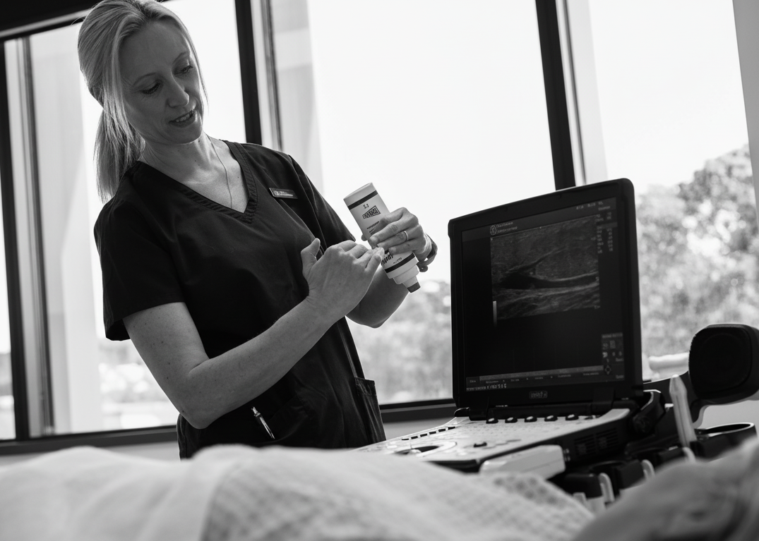
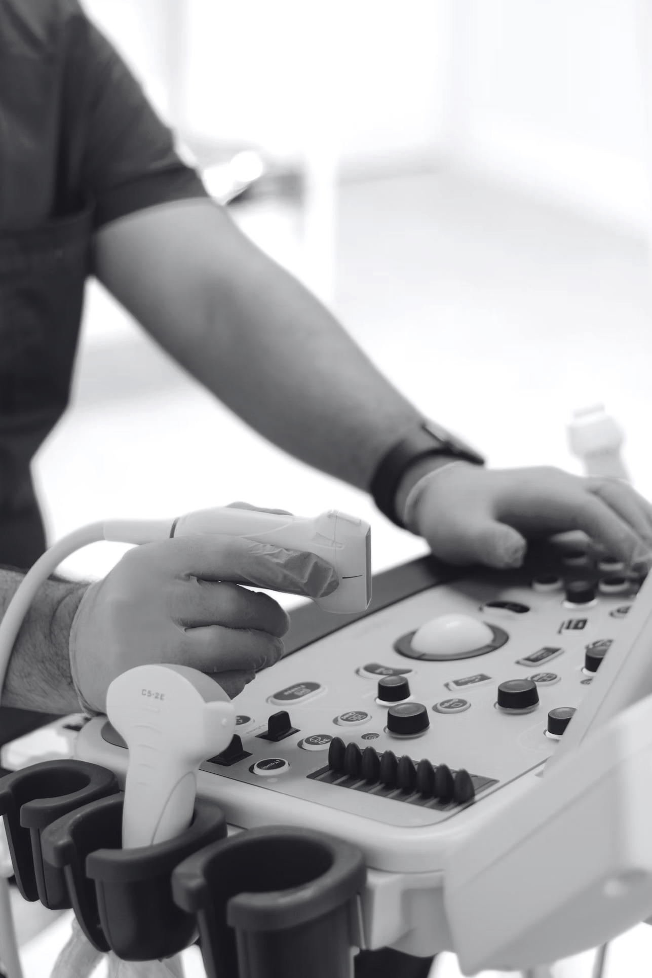
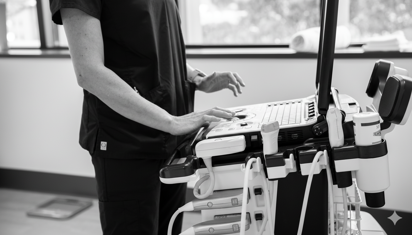
A vascular ultrasound uses high-frequency sound waves to create real-time images of arteries and veins, allowing our sonographers to assess blood flow, vessel walls, and surrounding tissues without radiation or injections.
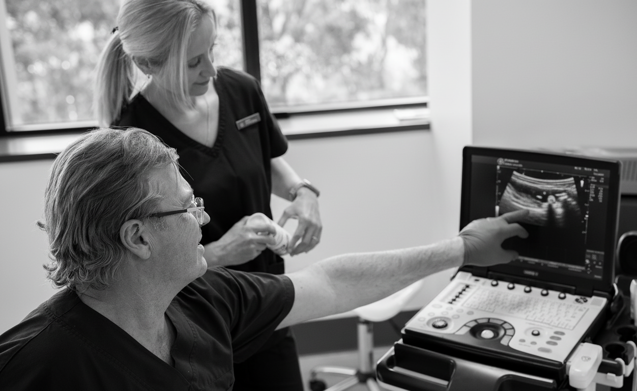
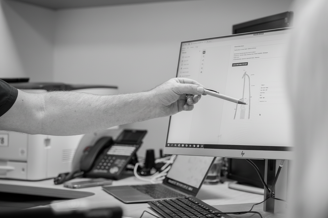
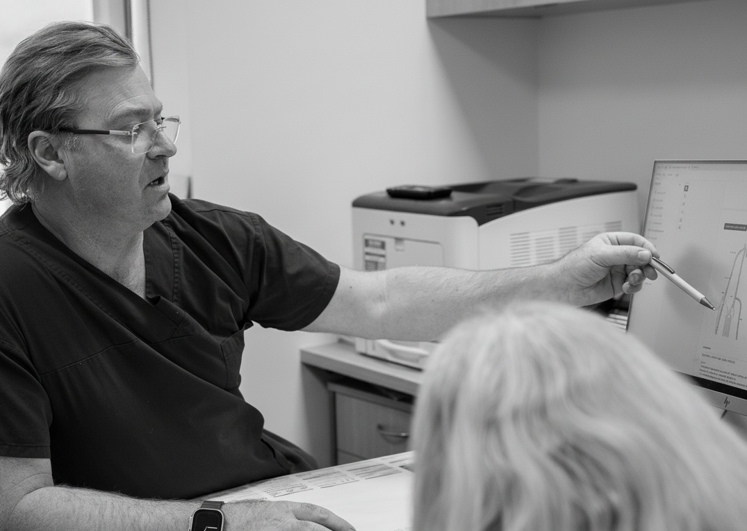
At Pulse Ultrasound every study is protocolled by vascular-trained clinicians, interpreted promptly, and shared securely with your referrer so treatment decisions can be made without delay.
Speak with our vascular team
Vascular Scan Overview
Browse the studies available in our dedicated vascular laboratory, then jump directly to the section that best matches your clinical question.
Carotid Arteries
A carotid duplex ultrasound visualises the common and internal carotid arteries, showing wall thickness, plaque, and the speed of blood moving toward the brain.
It is commonly requested for people with transient neurological symptoms, previous stroke, bruit on examination, or elevated cardiovascular risk, helping clinicians refine stroke-prevention plans.
Helps investigate
- Transient ischaemic attacks
- Visual disturbances or dizziness
- Monitoring known carotid plaque
Temporal Arteries
Temporal artery ultrasound is a fast, non-invasive way to look for the “halo sign” and thickening associated with giant cell arteritis, supporting or reducing the need for biopsy.
It is particularly helpful when patients develop new onset temporal headaches, scalp tenderness, jaw claudication, or acute vision changes.
Common reasons
- New persistent temple pain
- Sudden visual change
- Elevated inflammatory markers
Arm Arteries & Veins
This study reviews arterial inflow, venous patency, and the soft tissues of the upper limbs to detect narrowing, aneurysm, or thrombosis.
It supports investigation of arm swelling, numbness, cold extremities, and vascular access planning before dialysis or venous procedures.
Frequently requested for
- Swollen or painful arm
- Post-procedural follow-up
- Pre-access mapping

Thoracic Outlet Study
A thoracic outlet study examines the subclavian and axillary vessels while the arm is positioned in provocative postures, revealing vascular compression that can be missed at rest.
The assessment is useful for patients with positional arm heaviness, paraesthesia, venous engorgement, or suspected Paget-Schroetter syndrome.
When to book
- Symptoms triggered by overhead activity
- Suspected venous congestion
- Pre-surgical planning
Renal Arteries
Renal artery Doppler measures blood flow velocities to identify stenosis, aneurysm, or fibromuscular dysplasia affecting kidney perfusion.
It is recommended for treatment-resistant hypertension, unexplained changes in renal function, or surveillance of renal transplants.
Common indications
- Secondary hypertension workup
- Renal bruit on examination
- Transplant monitoring
Visceral Arteries
This scan evaluates the celiac, superior mesenteric, inferior mesenteric, and hepatic arteries to detect narrowing or aneurysmal change.
It can clarify causes of chronic abdominal pain, post-prandial discomfort, weight loss, or post-surgical follow-up needs.
Supports
- Chronic mesenteric ischaemia queries
- Postoperative surveillance
- Unexplained abdominal pain
Aorta & Iliac Arteries
Aortic and iliac ultrasound measures vessel diameters, looks for plaque or dissection flaps, and assesses flow into the lower limbs.
We routinely scan patients with pulsatile abdominal masses, family history of aneurysm, or to monitor previously repaired segments.
Highlights
- Maximum diameter measurements
- Endoleak surveillance
- Iliac inflow assessment
Abdominopelvic Veins
This evaluation follows the inferior vena cava, renal veins, gonadal veins, and pelvic venous plexus to detect obstruction, reflux, or compression syndromes.
It is valuable for investigating unexplained abdominal swelling, pelvic congestion, or atypical varicosities.
Useful for
- Pelvic congestion syndrome
- Nutcracker or May-Thurner queries
- Central venous obstruction
Varicose Veins
A comprehensive venous mapping identifies which superficial or perforator veins are contributing to reflux and visible varicosities.
The results guide vascular surgeons and phlebologists when planning surgery, laser therapy, or injectable treatments for aching, heavy, or cosmetic concerns.
Common symptoms
- Leg aching or heaviness
- Recurrent varicose veins
- Non-healing venous ulcers
Deep Vein Thrombosis (DVT)
DVT ultrasound uses compression and Doppler techniques to quickly detect blood clots in the deep veins of the legs or arms.
It is arranged urgently for acute calf pain, swelling, immobility, or post-operative and postpartum concerns, giving referrers clear guidance on anticoagulation.
Arrange promptly for
- Sudden limb swelling
- Calf pain after travel or surgery
- Elevated D-dimer
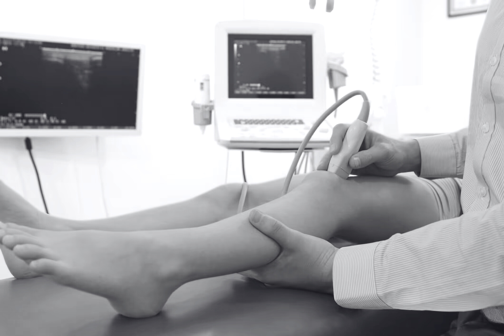
Leg Arteries & Veins
This assessment reviews femoral, popliteal, and distal arteries as well as venous return to diagnose peripheral arterial disease or chronic venous insufficiency.
It is useful for intermittent claudication, rest pain, delayed wound healing, or graft surveillance.
Highlights
- Duplex flow profiles
- ABI correlation
- Bypass graft checks
Vascular Malformations
High-resolution ultrasound differentiates low-flow venous malformations from high-flow arteriovenous lesions and documents their relationship to surrounding structures.
Imaging is recommended for congenital vascular anomalies, enlarging soft-tissue masses, or lesions that change with posture or activity.
Useful insights
- Flow dynamics and nidus mapping
- Tissue involvement
- Guidance for interventional planning
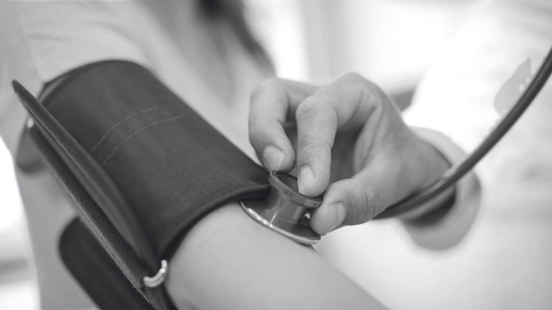
ABI (Ankle Brachial Index)
An ankle brachial index compares blood pressure at the ankle to the arm using Doppler probes, providing a simple ratio that screens for peripheral arterial disease.
It is often paired with toe pressures or duplex imaging for people with diabetes, smokers, and anyone with exercise-induced leg discomfort.
Why add ABI?
- Baseline before intervention
- Tracking treatment response
- Screening high-risk patients
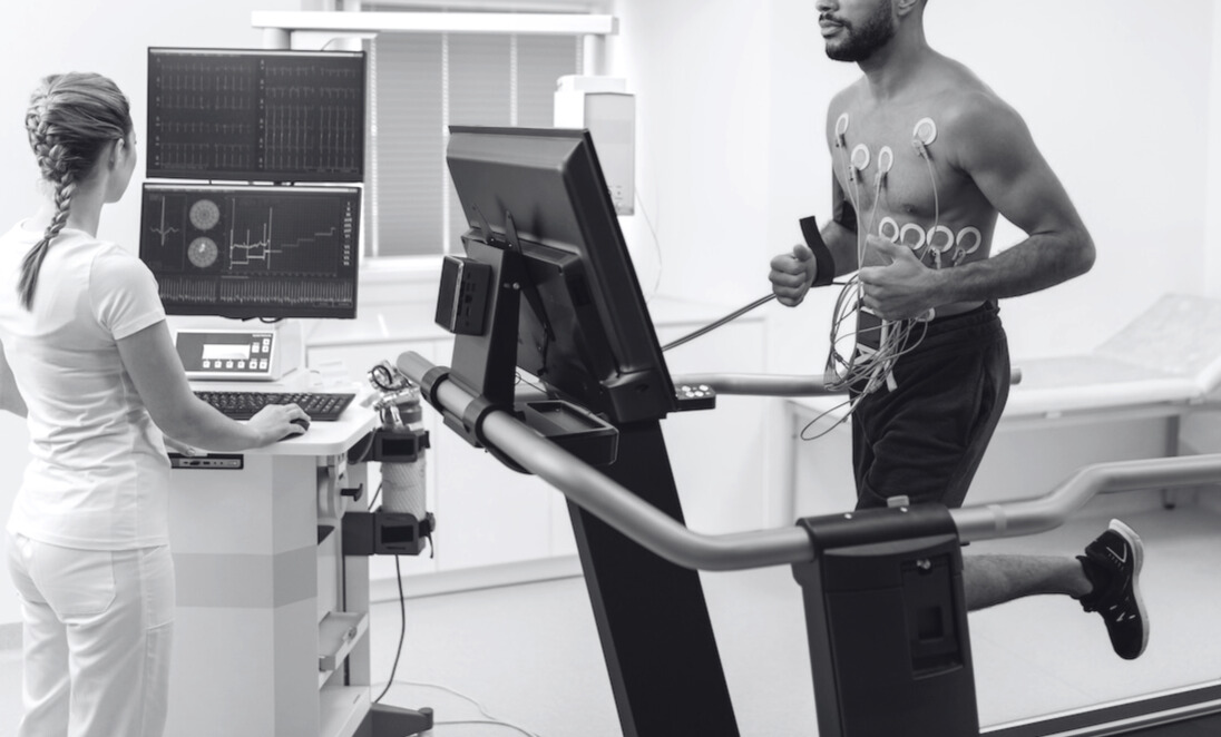
Exercise Treadmill Studies
Exercise testing evaluates blood flow before and after treadmill walking to unmask pressure drops or velocity changes that only occur with exertion.
It is ideal when symptoms such as calf tightness or numbness only arise during activity yet resting ultrasounds appear normal.
Ideal for
- Exercise-induced leg pain
- Chronic exertional compartment concerns
- Post-intervention benchmarking
Pelvic Congestion Studies
This targeted ultrasound visualises the ovarian and internal iliac veins to detect dilatation, reflux, or compression contributing to chronic pelvic pain.
It helps explain pelvic heaviness, vulvar or thigh varicosities, and discomfort that worsens after standing or pregnancy.
Helps with
- Chronic pelvic discomfort
- Perineal or vulval varices
- Postpartum venous symptoms
Make an Enquiry.
Request a callback or book your ultrasound scan. Referrals are welcome but not always required.
Phone: 0800 45 45 88
Email: info@pulseultrasound.nz
Office Address: 39 Ghuznee Street, Wellington, NZ